42 dog eye diagram
Browse 4,451 eye diagram stock photos and images available, or search for human eye diagram or eye diagram vector to find more great stock photos and pictures. Vintage anatomical color illustration of the muscles and bones of the face showing the eyeball and the circumorbital area. Diagram Of A Retinal Detachment. Here are a number of highest rated Hyper V Architecture Diagram pictures on internet. We identified it from obedient source. Its submitted by government in the best field. We resign yourself to this kind of Hyper V Architecture Diagram graphic could possibly be the most trending topic subsequently we allocation it in google improvement or facebook.
Differences Similarities. -less cone in the retina -Eyes located on the front of the skull. -Can only see small amount of colour -Same general structure. -Cant see stationery objects clearly. -Can see things clearer in motion. -Dogs can see in the dark. In the image above the top is what the human eye sees and the bottom is what the a dog sees.
Dog eye diagram
Jan 20, 2014 - Eye microdissection: the preclinical laboratory staff of Iris Pharma is trained for microdissection of ocular samples. Created Date: 6/11/2019 3:30:46 PM www.askmyvet.net
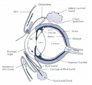
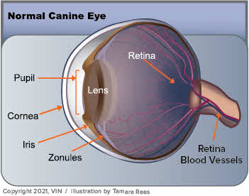
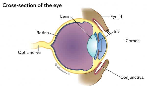
Dog eye diagram. A simple eye injury is one that penetrates or perforates the cornea or sclera of the eye – notice in the diagram above that these are injuries to the outer part of the eye. An example of a penetration would be a splinter piercing, whereas a perforation would be more like a scratch that goes across this part of the eye. The dog digestive system is very different from that of a human's, and it can take much longer for a dog to digest food. The dog digestion timeline can range from four to 10 hours, but perhaps even longer if he swallows a foreign object, which can lead to bowel obstruction in dogs. pepsininja. P. Samantha Avila. Random helpful things. Dog Anatomy. Treatment will depend on the specific type of eye abnormality that is affecting your dog. Depending on your veterinarian's experience with eye diseases, you may need further treatment with a trained veterinary ophthalmologist. Surgery can repair some congenital birth defects, and medicines can be used to mitigate the effects of some types of ... The dog has 321 bones. Regions of a Long Bone Structure of a Long Bone articular cartilage nutrient artery entering nutrient foramen marrow cavity compact bone spongy bone ligament periosteum endosteum physis (epiphyseal plate) physis (epiphyseal plate) metaphysis diaphysis metaphysis epiphysis epiphysis
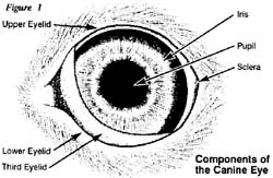
Arterial Blood Vessels of the Orbit. Page 5. General Anatomy. Dog. Blood Supply. Horizontal section. Long Posterior Ciliary a. Blood enters the globe. Short ... Jan 4, 2022 — The pupil is the black spot in the center of the eye. Dog pupils are round. The pupils should be the same size and should constrict to a ... Eye Structure and Function in Dogs - Dog Owners - Merck Veterinary Manual ... Canine ear anatomy Veterinary Care, Veterinary Medicine, Veterinary Technician ... Dog Eye Facts. A dog's eye functions much the same as any mammalian eye. The eyeball is round in shape with a light sensitive membrane, called the retina, lining the rear of the eyeball. Incoming light is focused and information is transmitted to the brain via the optic nerve. The dog's eye has a reflecting layer, called the tapetum lucidum ...
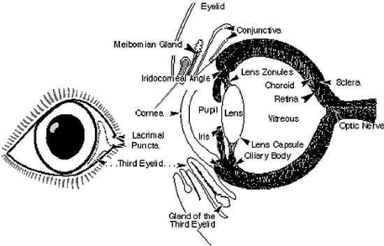
Special Senses of the Dog. The pictures in this section are reprinted with permission by the copyright owner, Hill's Pet Nutrition, from the Atlas of Veterinary Clinical Anatomy. These illustrations should not be downloaded, printed or copied except for personal, non-commercial use. Anatomy of the cat`s or dog`s eye. Royalty-Free Vector. Download preview. Anatomy of the cat`s or dog`s eye. Vertical section of the eye and eyelids. Third eyelid and Tapetum lucidum. Schematic diagram. detailed illustration. dog eye anatomy, cat anatomy, Oct 29, 2021 — Dog Eye Anatomy · Sclera: Tough, fibrous layer that's often referred to as the “white” of the eye · Cornea: Thin, clear layer at the front of the ... Aug 03, 2015 · Due to hundreds of years of selective breeding and the production by man of numerous types of dog breeds, dogs have the greatest variation of eye and orbit structure of any species. For example, such breeds as the brachycephalic dogs (those with short, wide heads) have eyes that appear to be more prominent, e.g. Pekingese, Boston terrier and pug.
Animal Eye Diagram High Resolution Stock Photography and ...
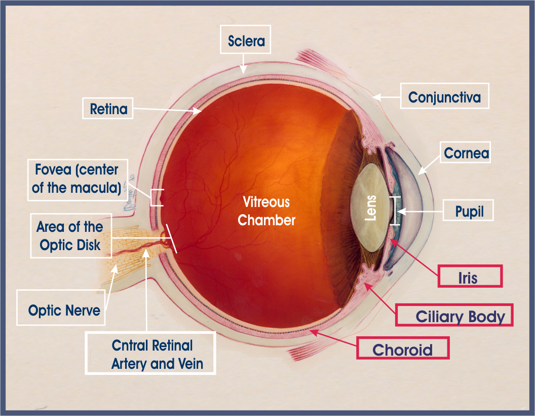
Eye Diagram Handout Author: National Eye Health Education Program of the National Eye Institute, National Institutes of Health Subject: Handout illustrating parts of the eye Keywords: parts of the eye, eye diagram, vitreous gel, iris, cornea, pupil, lens, optic nerve, macula, retina Created Date: 12/16/2011 12:39:09 PM

Dog Eye Anatomy Stock Illustrations – 52 Dog Eye Anatomy ...
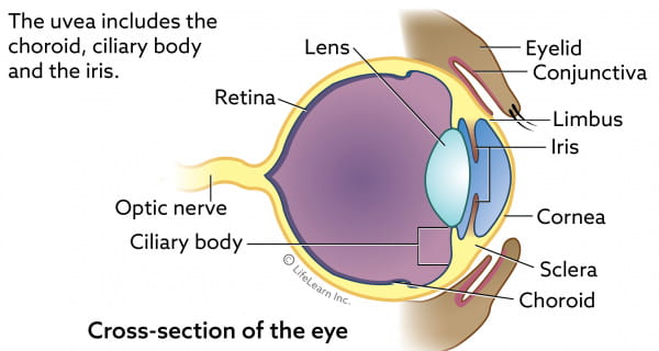
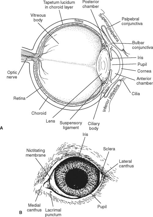
Functional Layers of a Dog Eye: On basis of function, the dog eye has three different layers: Fibrous Tunic: This part forms the outer most opaque layer and is comprised of collagen and fibrous tissues, called sclera. Sclera majorly forms the posterior of the eye, while anterior or the front part of the eye is the cornea, which is transparent and allows entry of light into the eye.

Eye Anatomy and Function in Animals | Eye anatomy, Dog eyes ...
The dog's eye is pretty much a garden-variety mammalian eye, with some notable adaptations that have evolved over the millennia.It is a globe with two fluid-filled chambers (anterior and posterior). The chambers are separated by the lens, the structure that helps focus light beams onto the rear part of the eye, the retina.
Your Pet's Eyes Are Gateways to the World: Are You Doing All ...
Apr 29, 2016 · Mark the page with two directional lines that divides the dog’s face in half. This helps with the symmetry of the face and lines up the eyes at one level. Pay attention to the fact that the face is round, not flat — the line marking the general position of the eyes is a curve. After that, put simple circles that define the eyeballs of the dog.

Anatomy of Dog with Inside Organ Structure Examination Vector ...
The Ophthalmic Exam. An ophthalmic exam is a thorough examination of the pet's eyes and the surrounding tissues. The exam may be performed by your veterinarian or by a veterinary ophthalmologist (an eye-care specialist). The exam is generally non-invasive and painless for your pet. The kind of tests performed depend on the nature of the pet ...
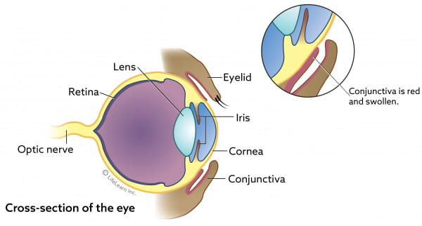
Conjunctivitis In Dogs | VCA Animal Hospitals
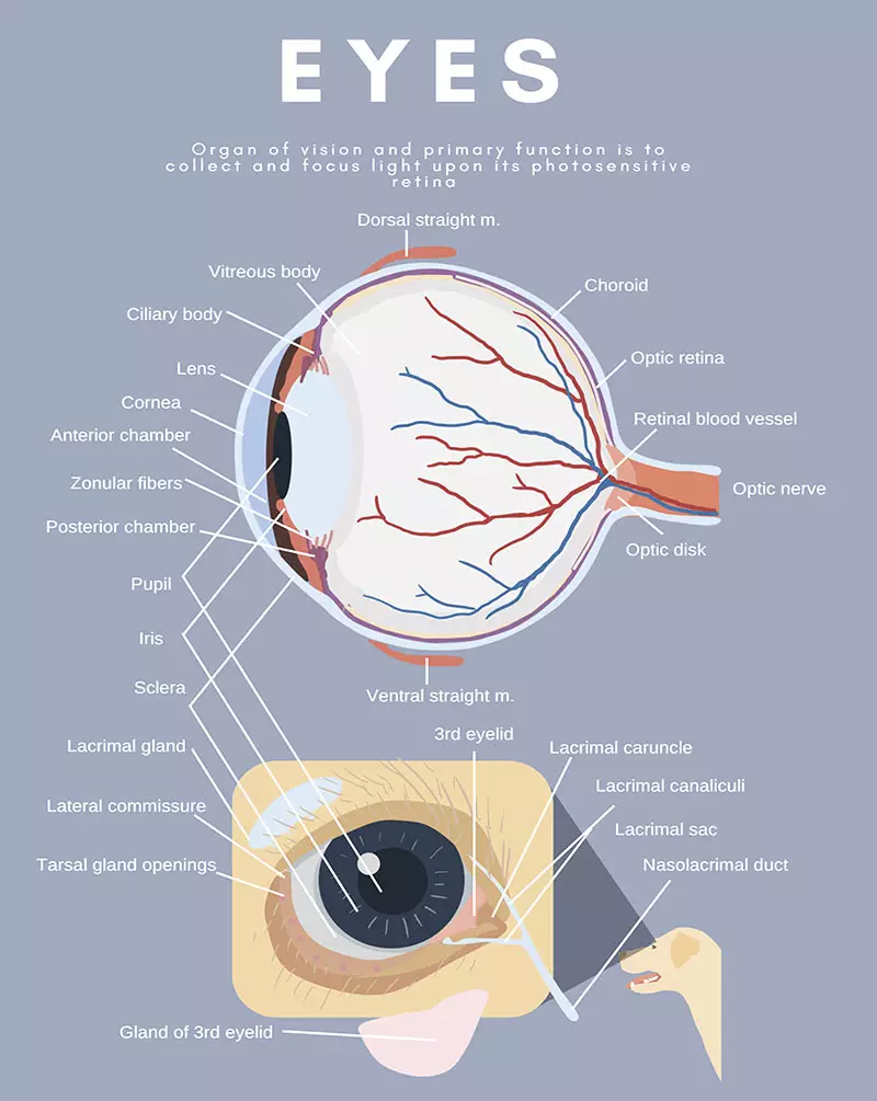
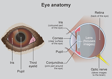
The eye is a paired organ, the organ of vision. The eye is made up of various components, which enable it to receive light stimuli from the environment, and deliver this stimuli to the brain in the form of an electrical signal. Vision involves all components of the eye. Structure. The eye is contained within the bony orbit of the head.

The Eyes: Window to your Dog's Health
Anatomy of the dog - Illustrated atlas This modules of vet-Anatomy provides a basic foundation in animal anatomy for students of veterinary medicine. This veterinary anatomical atlas includes selected labeling structures to help student to understand and discover animal anatomy (skeleton, bones, muscles, joints, viscera, respiratory system ...
Auckland Animal Eye Centre - Veterinary Eye Specialists - FAQ's
1. Your pooch may have been born without tear glands although this is very rare - it is more common is small breeds. 2. If your dog's third eyelid has to be removed then Dry Eye might develop. 3. If the tear glands have become damaged through injury or illness the this can cause the condition.

Observations in Ophthalmology: Corneal Opacities in Dogs & Cats
In a Dog's Eye. The visual system is an important sense available to the canine. ... Distichiasis is a condition in which small hair structures abnormally grow on the inner surface of the eyelids ( see diagram ). Both upper and lower lids may be involved. The abnormal hairs growing on the inner surface of the lids cause irritation to the cornea.
Jack Russell Terrier JRTCA: Medical - In a Dog's Eye
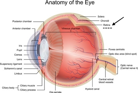
Vision is a complex phenomenon in which light emanating from objects in the environment is captured by the eye and focused onto the retinal photoreceptors (Figures 1-1 and 1-2).Electrical signals originating from these cells pass through a number of cell types in the retina and throughout the central nervous system (CNS) before arriving at the visual cortex, where the sensation of vision occurs.
Eye Tumors Melanoma In Dogs | VCA Animal Hospitals
The digestive system ( cat) ( dog) includes the mouth, teeth, salivary glands, esophagus, stomach, intestine, pancreas, liver and gall bladder. The digestive system absorbs and digests food and eliminates solid wastes from the body. The integumentary system is the skin and fur that cover the animal's body. The skin protects the underlying organs.

Ocular pharmacokinetics of atenolol, timolol and betaxolol ...
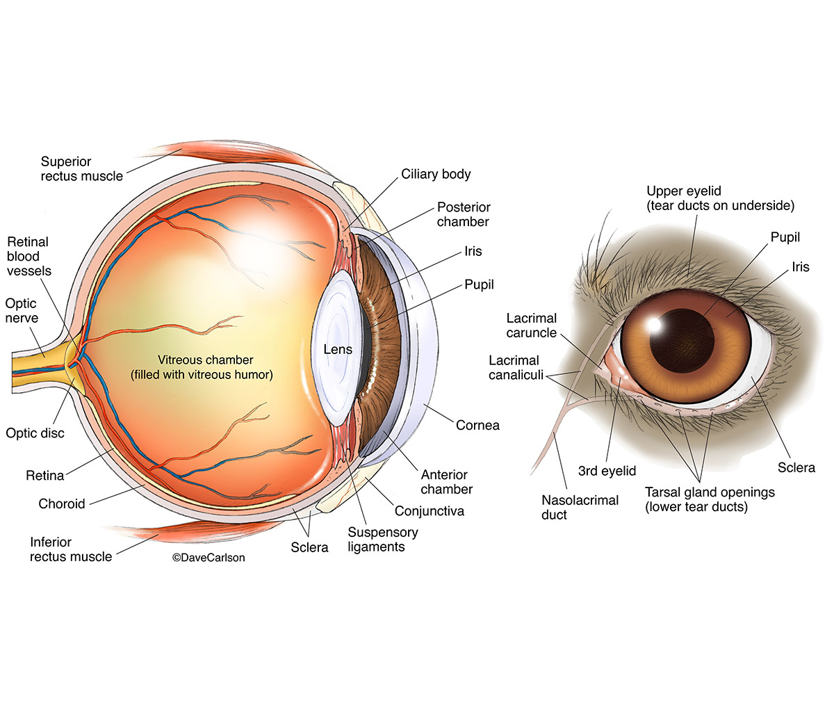
Anatomy of the eye. The bony cavity or socket that contains the eyeball is called the orbit. The orbit is a structure that is formed by several bones. The orbit also contains muscles, nerves, blood vessels, and the structures that produce and drain tears. The white of the eye is called the sclera. This is the relatively tough outer layer of the ...
Eye (ocular) anomaly in dogs and cats | Symptoms & Causes
Labeled anatomy of the head and skull of the dog on CT imaging (bones of cranium, brain, face, paranasal sinus, muscles of head) This module of vet-Anatomy presents an atlas of the anatomy of the head of the dog on a CT. Images are available in 3 different planes (transverse, sagittal and dorsal), with two kind of contrast (bone and soft tissues).

Behind Puppy Dog Eyes | WORLDkids
Dog's view. Visual acuity. Visual acuity is a measure of the spatial resolution of the visual system. It is often measured in cycles per degree (CPD), which measures how much an eye can differentiate one object from another in terms of visual angles. The maximum visual acuity of the human eye is around 50 CPD [7] and 60 CPD [8].

Normal eye anatomy Cornea: The cornea is transparent and more ...
Close-up face of Cute pug puppy dog sleeping rest open eye by chin. Border collie dog with different eye color. Picture showing a border collie dog with different eye color - blue/white and brown. Cute pomeranian dog smiling funny, with copy space, horizontal rectangular image, focus on the eye.

Eye Diagram Eyeball - Free vector graphic on Pixabay
The following diagram and paragraphs explain the skeletal anatomy of a dog. One extremely important part of a dog's skeletal anatomy is the skull. It is a long bone structure that encases the brain, and contains a cavity called the orbit, where the eye is located.

Dog eye anatomy explained | Brookfield Animal Hospital
www.askmyvet.net
Patient Guides - Uveitis.org | OIUF
Created Date: 6/11/2019 3:30:46 PM

File:Eye anatomy.jpg - Wikimedia Commons
Jan 20, 2014 - Eye microdissection: the preclinical laboratory staff of Iris Pharma is trained for microdissection of ocular samples.
Eye Structure and Function in Cats - Cat Owners - MSD ...

Primary Lens Luxation – Chevromist Puppies Australia

Corneal Ulcer in Dogs - Causes, Symptoms and Treatment

Manage Your Dog's Eye Problems Naturally - Dogs Naturally ...
Anatomical, histological and computed tomography comparisons ...
VETzInsight - VIN
Dog Eye Anatomy | Carlson Stock Art
Page 2 - Animal Eye Diagram High Resolution Stock Photography ...

Eye Health: A Foreign Body in the Eye — Eyes! on Broadway
Eye problems in dogs - PDSA

Manage Your Dog's Eye Problems Naturally - Dogs Naturally ...

Components of human eye. Illustration about Anatomy and ...

Vector Stock - Diagram of human eye anatomy with label. Stock ...

Canine Eye (fluid) | Annette's Vet Student Info
Diseases of the Eye | Veterian Key
Lenticular Sclerosis In Dogs | VCA Animal Hospitals
Progressive Retinal Atrophy in Dogs (PRA)

Dog Eye Care - Cynology Hub: mygsdorg

Dog eye anatomy at Little Critters vet | Vet medicine, Animal ...
Third Eyelid | Veterian Key

Photographs of the right eye and eyelids in a representative ...

An introduction to cataracts | The Veterinary Nurse

The anatomy of a dogs eye | Pets and Animals


Comments
Post a Comment