42 enzyme diagram unlabeled
Download scientific diagram | Relaxation activity of CPM labeled MtTOP1-K524C mutant. Serial dilutions of unlabeled MtTOP1-K524C mutant and CPM labeled MtTOP1-K524C mutant proteins were assayed ... Posted above is the anatomical diagrams of the digestive system. The human digestive system consists of the gastrointestinal tract plus the accessory organs of digestion (the tongue, salivary glands, pancreas, liver, and gallbladder). In this system, the process of digestion has many stages, the first of which starts in the mouth. Most of the digestive organs are tube-like and contain the food ...
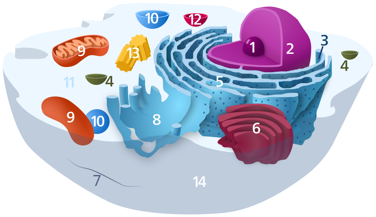
Human Cell Diagram, Parts, Pictures, Structure and Functions The cell is the basic functional in a human meaning that it is a self-contained and fully operational living entity. Humans are multicellular organisms with various different types of cells that work together to sustain life.

Enzyme diagram unlabeled
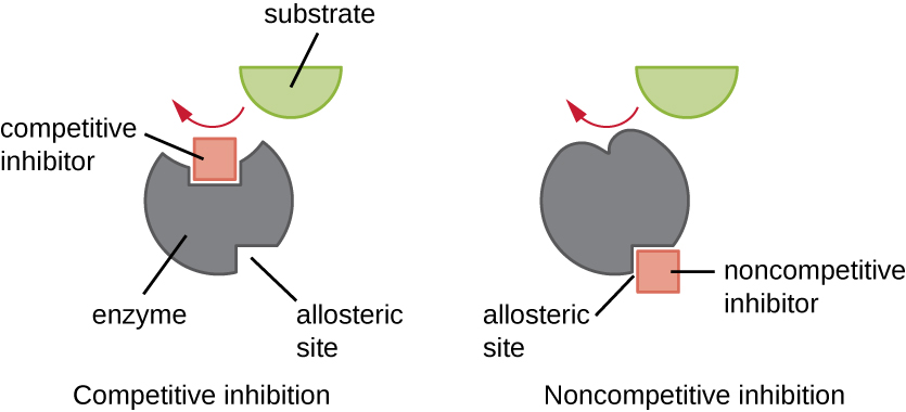
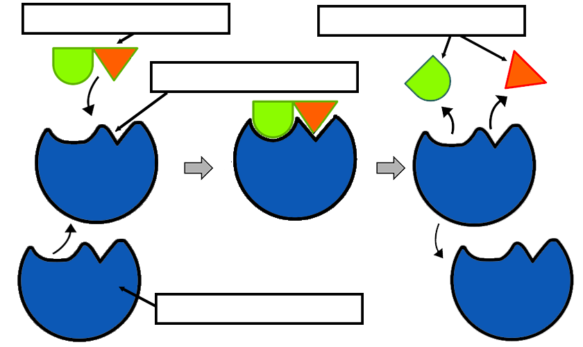
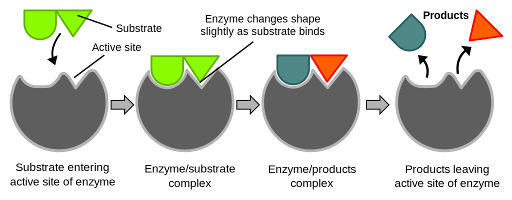
Introduction - Enzyme Characteristics: The basic mechanism by which enzymes catalyze chemical reactions begins with the binding of the substrate (or substrates) to the active site on the enzyme. The active site is the specific region of the enzyme which combines with the substrate. The binding of the substrate to the enzyme causes changes in the distribution of electrons in the chemical bonds ... Glycolysis is a process in which glucose divided into two pyruvate molecules. However, it is assumed as a linear pathway of ten enzyme meditation steps. This pathway has two stages or phases; the energy investment phase and the energy generation phase. In it, the first five steps out of ten are Energy Investment Phase or preparatory phase that ... There is an unlabeled diagram in the end of the article for readers to practice labeling. Tea Processing Flow Chart. In the stomach acid pepsin mucus and lipase enzymes are released. There are four primary stages of food digestion in the human body that include.
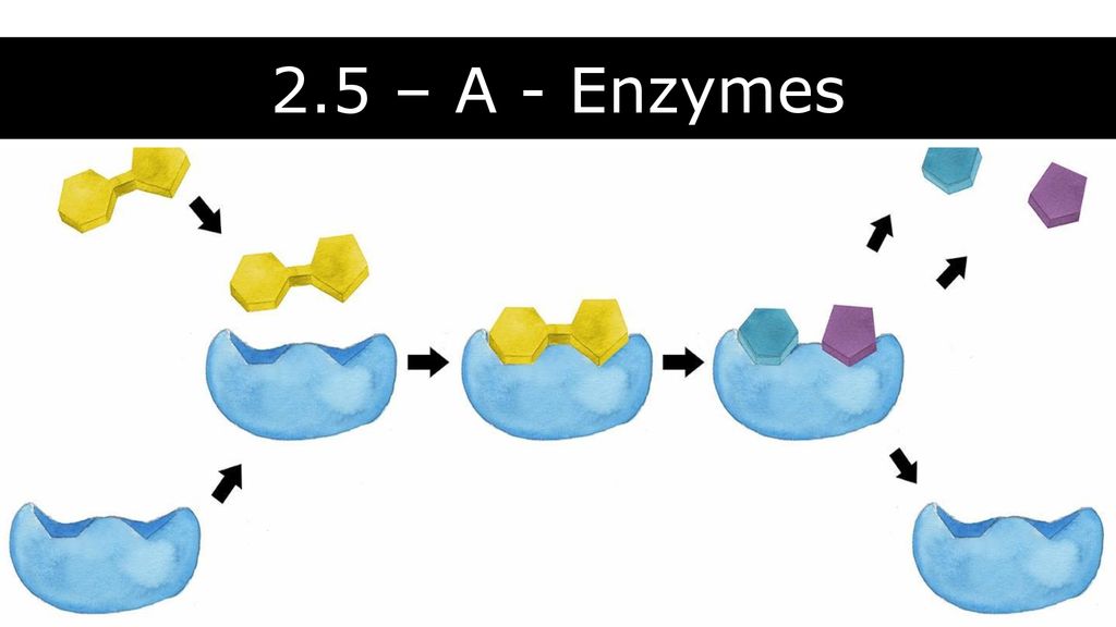
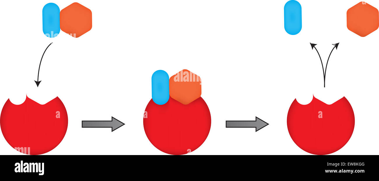
Enzyme diagram unlabeled. You may wish consider printing the unlabeled diagrams, and labeling the significant structures yourself as you read or as diagrams are discussed in class. As an AP student, you are expected to be an active participant, so bring all diagrams to class for every unit. ... 8.15: The effect of an enzyme on activation energy 8.17: The active site and ... Immunoassays are used to quantify molecules of biological interest based on the specificity and selectivity of antibody reagents generated. In HTS and lead optimization projects, assays are designed to detect molecules that are produced intracellularly or secreted in response to compounds screened. This chapter describes the basics of designing and implementing robust, automation friendly ... The diagram shows how this works. In this example, the enzyme splits one molecule into two smaller ones, but other enzymes join small molecules together to make a larger one. Labeled and unlabeled analyte are present in equal amounts. c. The concentration of patient analyte is inversely proportional to bound label. d. All the patient analyte is bound in the reaction. ... Enzyme activity is altered when binding to antibody occurs. b. The enzyme label is on the antibody.
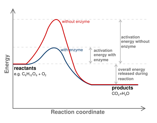
Enzyme Diagram Label: Substrate, Enzyme, Active Site, Free Energy, Progress of the Reaction, Enzyme-Substrate Complex, Products Reactants, Products, With Enzyme, Without Enzyme, Free Energy of Activation Enzyme Diagram ... Human digestive system diagram unlabeled 25. Digestive System Infections Describe the major anatomical features of the human digestive system Describe the normal microbiota of various regions in the human digestive system Explain how microorganisms overcome the defences of the digestive tract to cause infection or intoxication Describe general signs and symptoms associated with You may be offline or with limited connectivity. ... Download Skeletal system diagram worksheet body systems chart worksheet and unlabeled body cavity diagram are three of main things we want to show you based on the post title. Anatomical terminology for body cavities. Anatomy Introduction To Anatomy Biology College Medical Terminology Study Biology Activity In addition to the major body cavities the body also contains […]
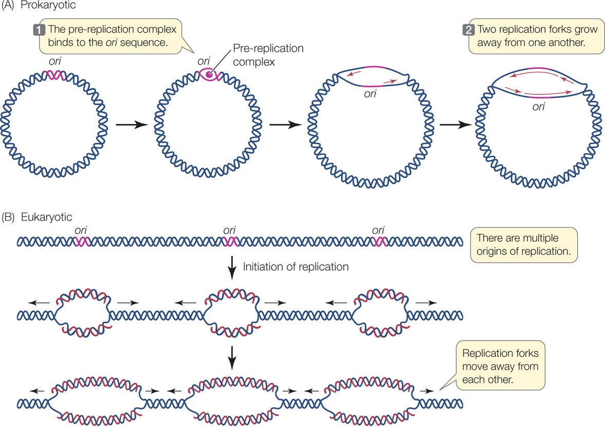
Labeled Pfizer confidential the study is known as a bio-distribution study that uses luciferase enzymes and radioisotope markers to accurately track the distribution of Pfizers mRNA LNPs across the body. The first section is labeled. There is an unlabeled diagram in the end of the article for readers to practice labeling. 1627x1606 Digestive Enzymes Diagram Unlabeled - Sketch Of Human Digestive System. 0 14. 304x429 Diagram Volcano (Unlabeled) - Volcano Sketch. 0 3. 1200x927 Printable Plant Cell Diagram Labeled, Unlabeled, And Blank - Plant Cell Sketch. 0 3. 800x800 Stack, Pile Of Shiny Blank, Unlabeled Gold Coins, Sketch Vector - Coin Sketch. 0 0. The diagram below shows the structure and functions of the human digestive system. Let learn the different parts of the human digestive system. Mouth — It includes teeth, salivary glands and tongue. It is the beginning of the digestive tract and the process of digestion begins from the mouth, where teeth help by breaking and grinding the food ... DNA and completely unlabeled DNA are found. When two generation times have elapsed after the addition of 14N, half-labeled and unlabeled DNA are present in equal amounts." A conservative mode of replication is ruled out by the observation that all the DNA formed a band
Enzyme Substrate Complex An Overview Sciencedirect Topics
nephron diagram unlabeled - Google Search Kidney Anatomy, Medical Anatomy, Renal Physiology, Handwritten Tutorials, renal function of a nephron.Annotate a diagram of a Note: The diagram above is a A. Overview Diagram Diagram Nephron Diagram Nephron, diagram nephron diagram. alt. Nephron Diagram Blank is free HD wallpaper.

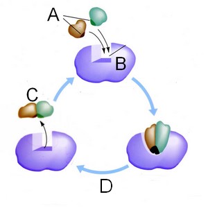
The Normal Shape Of An Enzyme Is As Shown In Structure A If The Enzyme S Shape Changes To That Shown Brainly Com
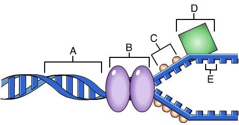
During DNA synthesis, nucleotides are added to the 3 end of the growing new strand—the end at which the DNA strand has a free hydroxyl (—OH) group on the 3′ carbon of its terminal deoxyribose.In summary, the DNA template is read 3′ to 5′, while the new strand of DNA is generated 5′ to 3′, forming an antiparallel double helix.. As we noted in Concept 3.1, a free nucleotide can ...
Mrhendersonatrampart Weebly Com
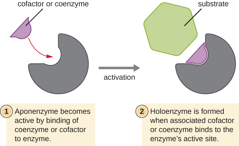
Enzyme Substrate Complex Definition. The enzyme substrate complex is a temporary molecule formed when an enzyme comes into perfect contact with its substrate. Without its substrate an enzyme is a slightly different shape. The substrate causes a conformational change, or shape change, when the substrate enters the active site.The active site is the area of the enzyme capable of forming weak ...
Intro To Cell Bio Chapter 1
Talking related with Unlabeled Digestive System Diagram Worksheet, we have collected some similar photos to add more info. small intestine digestive system diagram unlabeled, diagram of human body glands endocrine and human digestive system diagram without labels are three of main things we want to present to you based on the gallery title.

Biology Pap Enzyme Structure Function Quiz Flashcards Quizlet
Label the organs of digestive system. Food is broken down in the digestive system. Help children to cut the different human body organs and place them in the right place on the body template. Potassium is a mineral thats crucial for life. There is an unlabeled diagram in the end of the article for readers to practice labeling.
1
Diagram 2 • Show the correct enzyme at the initiation spot (pick one end) • Show unzipping • Show the mRNA - 15 bases long (You want to have a start codon at the beginning of the mRNA and an end a stop codon at the end of the mRNA) • Show how mRNA is different from DNA by using a different color

Enzyme Molecule An Overview Sciencedirect Topics
The enzyme phosphoglycerate kinase transfers the high-energy phosphoryl group from the carboxyl group of 1,3-bisphosphoglycerate to ADP, forming ATP and 3-phosphoglycerate. This is a unique example where ATP can be produced at substrate level without participating in electron transport chain.
Topic Enzymes Ppt Download
Diagram of the Digestive System And an Explanation of its Working. Digestive system helps in breaking complex food into simpler forms. With the help of a diagram in this article, let us understand the function of this system, and the organs that constitute it. There is an unlabeled diagram in the end of the article for readers to practice labeling.
Immunoassay Wikipedia
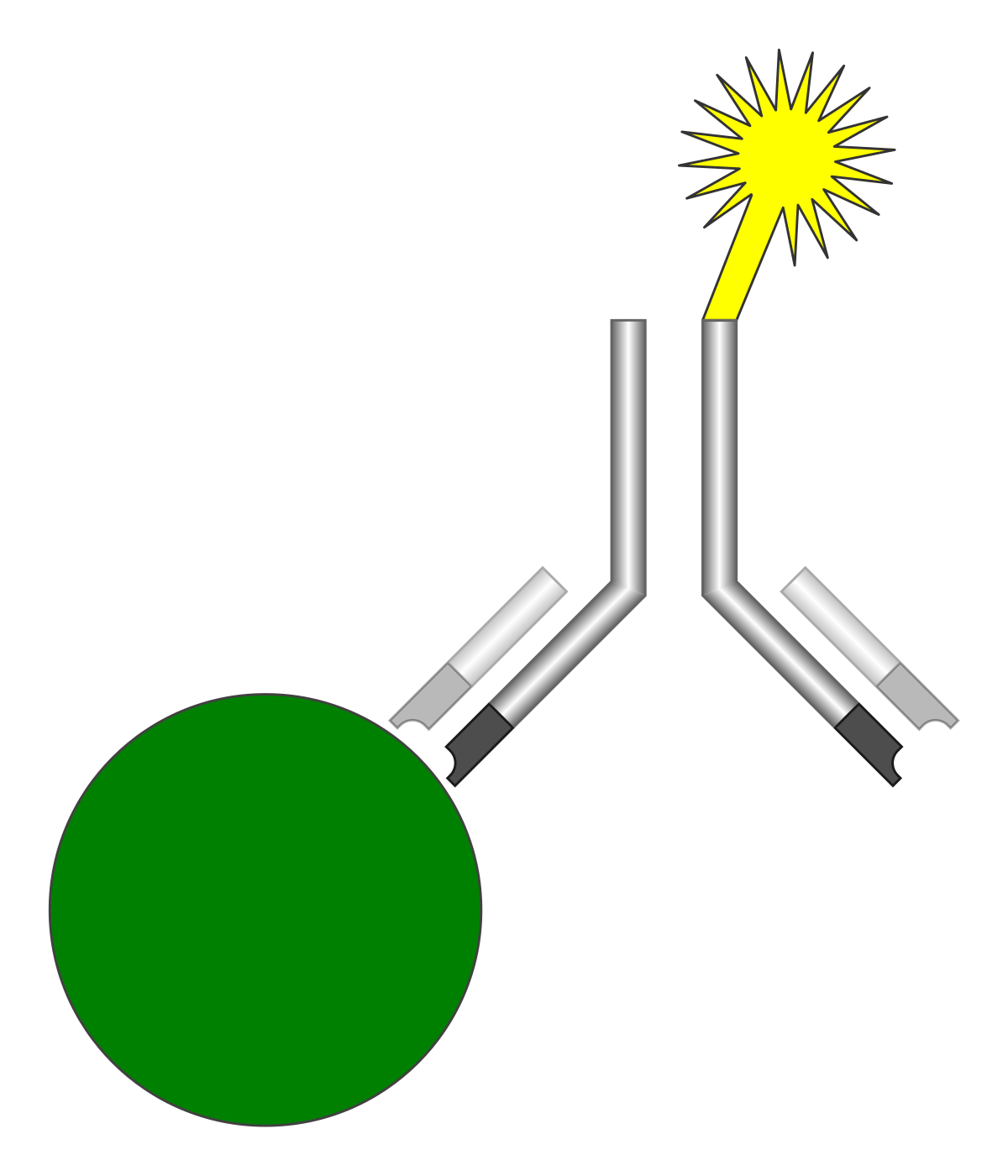
Enzyme-labeled antibodies and corresponding chromogen substrates (chromogenic detection) ICC/IF Cells Fluorochrome-labeled antibodies (immunofluorescent detection) IF Tissues or Cells Immunofluorescent detection, can be applicable to ICC and/or IHC Figure 1. Examples of staining using traditional ICC, ICC/IF, and IHC with IF based detection

The Big 4 Macromolecules Ppt Download
There is an unlabeled diagram in the end of the article for readers to practice labeling. Tea Processing Flow Chart. In the stomach acid pepsin mucus and lipase enzymes are released. There are four primary stages of food digestion in the human body that include.
Hillis2e Ch09
Glycolysis is a process in which glucose divided into two pyruvate molecules. However, it is assumed as a linear pathway of ten enzyme meditation steps. This pathway has two stages or phases; the energy investment phase and the energy generation phase. In it, the first five steps out of ten are Energy Investment Phase or preparatory phase that ...

Quia 9ap Chapter 8 An Introduction To Metabolism Detailed
Introduction - Enzyme Characteristics: The basic mechanism by which enzymes catalyze chemical reactions begins with the binding of the substrate (or substrates) to the active site on the enzyme. The active site is the specific region of the enzyme which combines with the substrate. The binding of the substrate to the enzyme causes changes in the distribution of electrons in the chemical bonds ...
1
8 1 Energy Redox Reactions And Enzymes Microbiology Canadian Edition

Enzymes And Biological Reactions Flashcards Quizlet

Large Scale Enzyme Based Xenobiotic Identification For Exposomics Nature Communications
Analyzing Graphics Enzymes

Which Diagram Most Correctly Represents The Process Of Mitosis

Scheme Of The Electrochemical Enzyme Linked Dna Sensing Procedures A Download Scientific Diagram
1

Enzyme Substrates An Overview Sciencedirect Topics
Lock And Key Mechanism Of Enzymes Stock Photo Alamy

Enzyme Linked Immunosorbent Assay Elisa A Practical Tool For Rapid Diagnosis Of Viruses And Other Infectious Agents Semantic Scholar

Enzymes Warm Up Th And Fri Science Quizizz
Enzymes Biology For Majors I
Mastering Biology Chapter 16 Rhs Homework

Detection Of Nitrocefin Recyclization Based On Competitive Acylation Of Download Scientific Diagram

Enzyme Label Diagram Quizlet

Hsc Biology Maintaining A Balance Notes Dot Point Summary Dux College
Analyzing Graphics Enzymes
Lysosome Wikipedia

Principle Of The Alphascreen Cgmp Pde Assay The Cgmp Pde Enzyme Download Scientific Diagram
8 1 Energy Redox Reactions And Enzymes Microbiology Canadian Edition
Chapter 8 Enzymes

Epr Spectra Of Wild Type And Isotopically Labeled Anaerobic Download Scientific Diagram

If40kmhw0gam5m

Biology Notes For A Level 22 Summary Of Enzymes
3dmoleculardesigns Com
Enzymes Biology For Majors I

The Unlabeled Antibody Enzyme Method Of Immunohistochemistry Preparation And Properties Of Soluble Antigen Antibody Complex Horseradish Peroxidase Antihorseradish Peroxidase And Its Use In Identification Of Spirochetes Semantic Scholar
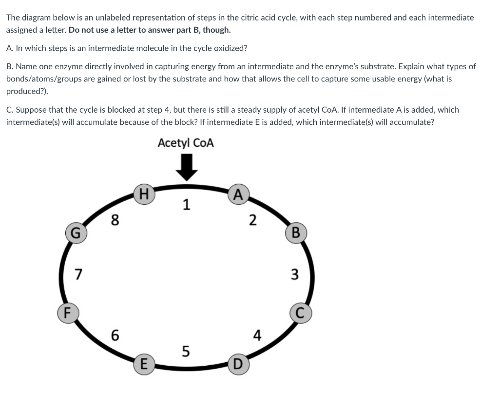
Solved The Diagram Below Is An Unlabeled Representation Of Chegg Com
Quiz Enzymes


Comments
Post a Comment