38 diagram of villi
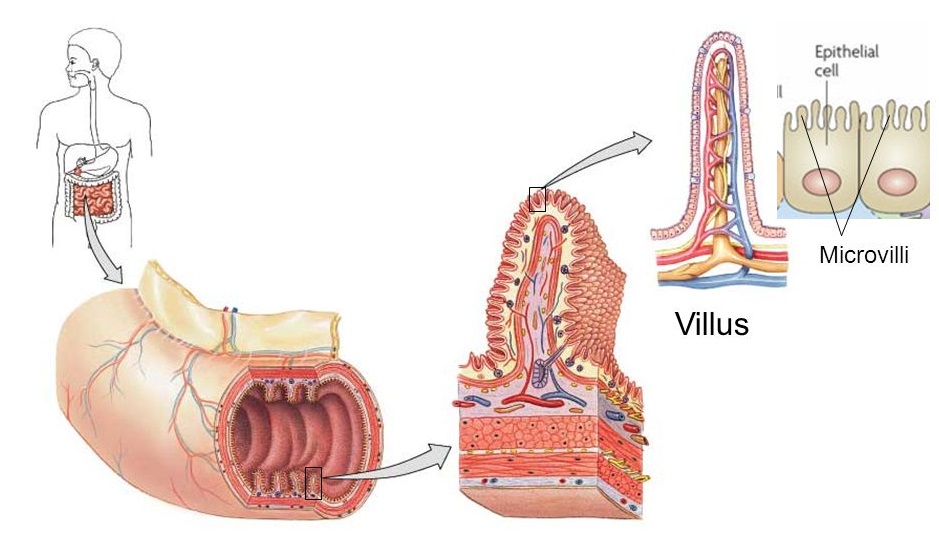
villus, plural villi, in anatomy any of the small, slender, vascular projections that increase the surface area of a membrane.Important villous membranes include the placenta and the mucous-membrane coating of the small intestine.The villi of the small intestine project into the intestinal cavity, greatly increasing the surface area for food absorption and adding digestive secretions. Biology. High School. answer. answered. Look at the diagram of villi. Which label points to a capillary? 2. See answers. report flag outlined.
Best viewed on 1280 x 768 px resolution in any modern browser. Villi diagram 343. Villi diagram 344. Villi diagram 352. Villi diagram 358. Villi diagram 370. Villi diagram 387. Villi diagram 392. Villi diagram 402.

Diagram of villi
c. Renewal of the layers in contact with the villi which helps in absorption. 2. Propulsion: The chyme is moved over the large area of small intestine to facilitate digestion and absorption and the residues are propelled downwards to the ileocecal junction to reach the large intestine, mostly for excretion. Find villi definition, villi location, and villi function. Learn the structure of villi, and the role and purpose of villi in the small intestine. Updated: 09/16/2021 The villi are small finger-like projections of the wall of the small intestine which extend into the lumen or interior space of the small intestine. As digestion is completed in the small intestine, the villi are then bathed in a fluid which contains the nutrient subunits the cells need. These subunits include monosaccharides, amino acids ...
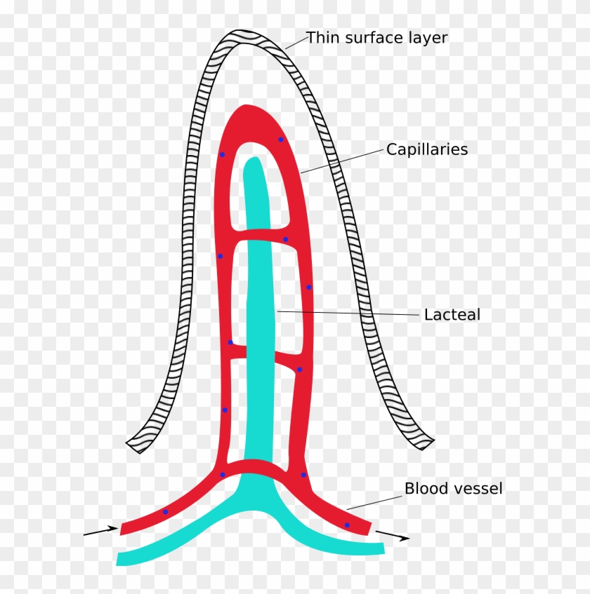
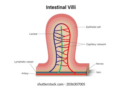
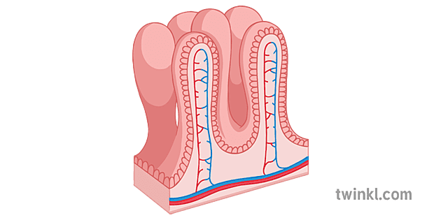
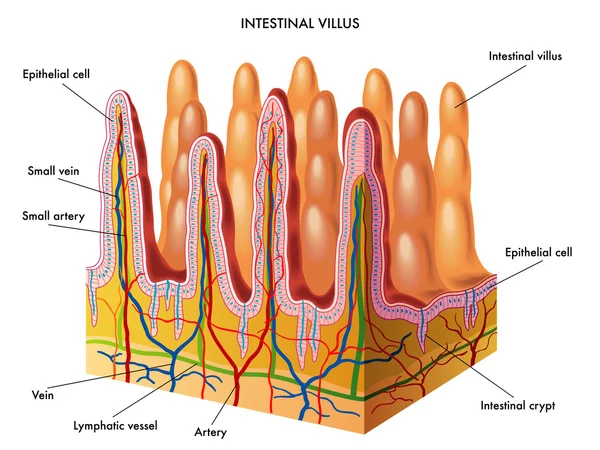
Diagram of villi. Villi. These Villi tiny are fingerlike projections through which the nutrients are absorbed into the bloodstream. The Villi capture nutrients as they move through the small intestine. Villi Photograph of Villi magnified (very high power) Note; your microscope will not show nearly the detail as in this picture. Blood vessels Glands secreting Villi in the small intestine absorbs nutrients and completes the breakdown of food. Factors of its structure that help it function include. The process that the nutrients move into the villi is diffusion. The picture above is a diagram of what is inside the villus. It explains what kind of nutrients is absorbed by the blood capillary which is ... Start studying Movement of Nutrients via Villi. Learn vocabulary, terms, and more with flashcards, games, and other study tools. Virtally all nurtients, including all amino acids and sugars, enter the body across the epithelium covering small intestinal villi. As shown in the diagram above, each villus contains a capillary bed and a blunt-ended lymphatic vessel referred to as the "central lacteal".
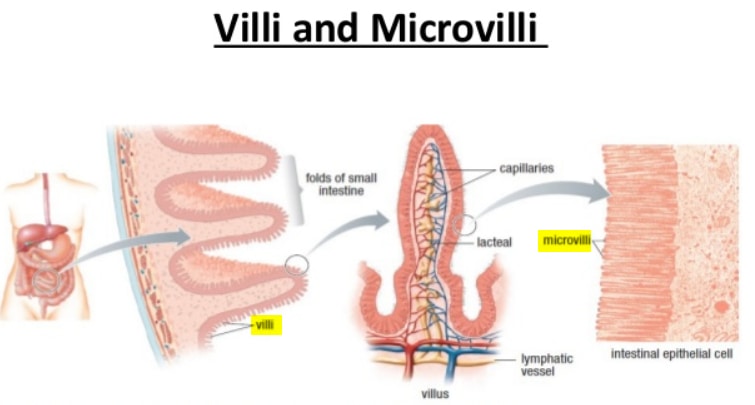
The main difference between villi and microvilli is that villi are small projections on the mucous membrane, particularly in the small intestine. But, microvilli are tiny extensions that mainly occur on the cell membrane of different organs. Furthermore, villi increase the surface area while microvilli are responsible for absorption and secretion, increasing the surface area. Villi Diagram. Below given picture is the villi diagram: [Image will be uploaded soon] Role of Villi. The role of villi or villi function are stated below: The villi along with the microvilli support in increasing the intestinal adsorbent surface area by approximately 30-fold and 600-fold, respectively. Sep 20, 2018 - villi diagram simple kidney diagram bladder diagram parts of a villus small intestine crypts. Villi is the plural of villus. The epithelial cells that cover each villus themselves have projections called microvilli. A close up of the villi in the small intestine Cross-section of a villus.
Draw a schematic diagram of villus in small intestine. Explain how digestive system coordinate with circulatory system. (AS5) Draw the diagram of villi in small intestine and label its parts. Medium. View solution > In an adult human, the length of the small intestine is about. Medium. View solution > Villi are present in _____. Medium. View solution > View more. More From Chapter. Digestion and Absorption. View chapter > Shortcuts & Tips . HOW to draw VILLI Easily. Easy Drawing Step by Step for students.Villi increase the internal surface area of the intestinal walls making available a greater ... How to draw structure of Villi step by step for class Student in easy way. Any student can draw this villi Diagram easily.
Intestinal Villi Diagram Royalty Free Vector Image
Functions. Their function is to increase the surface area of the small intestinal wall for absorption of the digested food. These projections absorb the protein molecules and help in the transfer of the proteins to all cells and tissues. Many blood vessels are present within these villi, that help in the absorption of digested food and carry it ...
Intestinal Villus Simplified Labeled Diagram Of A Villus Free Transparent Png Clipart Images Download
Draw the diagram of villi in small intestine and label its parts. Biology. Q2. Identify the part labelled [x] in the diagram. Biology. Q3. (a) Write the function of following parts in human female reproductive system : (i) Ovary (ii) Oviduct (iii) Uterus
Microvilli Definition Diagram Vs Villi And Cilia And Function Laboratoryinfo Com
The diagram provided shows the villi that line the ileum of the small intestine. What is the main purpose of the villi? A To trap pathogens, dust, and other unwanted substances and move them toward the stomach to be broken down; B To decrease ...

Villus
Villi - Small Intestine. Create healthcare diagrams like this example called Villi - Small Intestine in minutes with SmartDraw. SmartDraw includes 1000s of professional healthcare and anatomy chart templates that you can modify and make your own.
Draw A Neat Labelled Diagram Of A Section Of Small Intestinate Mucosa Showing Villi Sarthaks Econnect Largest Online Education Community
2. The Villus: The villus is a slender finger-like fold of the mucous membrane projecting outward from the surface. The surface of the mucous membrane is covered with numerous such projections and the structures of villi in the different parts of the small intestine are leaf-shaped in the duodenum, rounded in the jejunum and club-shaped in the ileum.

Intestinal Villi Cross Section Of The Enlarged Part Of The Duodenum Full Color Diagram Stock Vector Adobe Stock
Intestinal villi (singular: villus) are small, finger-like projections that extend into the lumen of the small intestine.Each villus is approximately 0.5-1.6 mm in length (in humans), and has many microvilli projecting from the enterocytes of its epithelium which collectively form the striated or brush border.Each of these microvilli are about 1 µm in length, around 1000 times shorter than ...

Chapter 18 The Small And Large Intestines Bio 140 Human Biology I Textbook Libguides At Hostos Community College Library
SINGLE VILLI : Intestinal villi (singular: villus) are small, finger-like projections that extend into the lumen of the small intestine. Each v...

Villi Diagram Quizlet
Villi. Create healthcare diagrams like this example called Villi in minutes with SmartDraw. SmartDraw includes 1000s of professional healthcare and anatomy chart templates that you can modify and make your own.

Villi And Microvilli Of Small Intestine Diagram Quizlet
Villi diagram. Villi Create healthcare diagrams like this example called Villi in minutes with SmartDraw. SmartDraw includes 1000s of professional healthcare and anatomy chart templates that you can modify and make your own. 70/71 EXAMPLE The villi are small, finger-like projections about a millimeter in length that protrude from the circular folds.

The Diagram Given Below Represents A Section Of Small Intestinal Mucosa Identify A B And C Img Src Https D10lpgp6xz60nq Cloudfront Net Physics Images Ncert Fing Bio Obj Xi Da C16 E01 012 Q01 Png Width 80
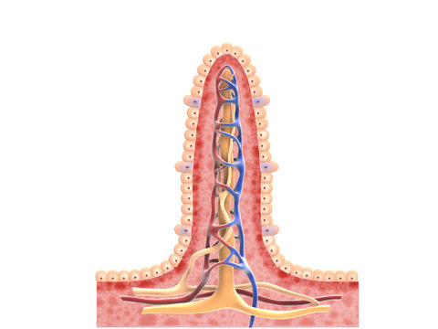
This diagram represents the types of bone-marrow cell derivatives operative within the lamina propria. They include (in cerise) the subepithelial myofibroblast system (MYF); pericytes (green) supporting the subepithelial capillaries and main vasculature of the villi (artery, red: vein, blue); the lacteal (L) supported by smooth muscle cells (SM) and (purple) the muscularis mucosae (MM).

Intestinal Villi Bioninja
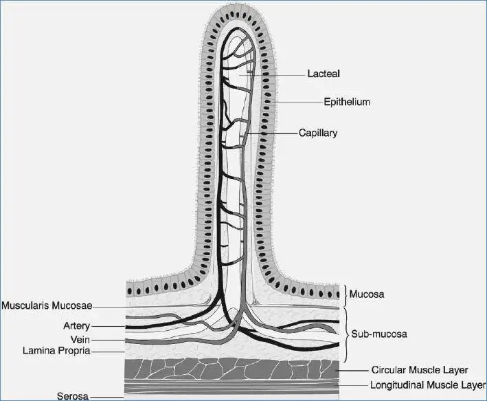
The villi are tiny hair-like projections of the intestinal surface into the lumen of the intestine. These structures increase the available surface area of the ...
Ib Biology Sl Digestion
Absorption is the movement of digested food molecules through the wall of the intestine into the blood or lymph. The small intestine is the region where digested food is absorbed. Most absorption ...
2 443 Best Villi Images Stock Photos Vectors Adobe Stock
The lobular arrangement of villi . Vascular cast showing fetal vasculature in white and the maternal arterial blood in red . Wigglesworth • The villi are arranged into a series of 30- 40 lobules, each centred over the opening of a spiral artery • Each lobule acts as an independent maternal -fetal exchange unit
What Would Be The Result If There Were No Villi In The Intestinal Tract And The Walls Of The Intestines Were Smooth Socratic
11 Sept 2017 ... The transfer of food particles from the digestive system to the circulatory system takes place at the inner lining of the small intestine, ...
Villi Images Stock Photos Vectors Shutterstock
The villi are small, finger-like projections about a millimeter in length that protrude from the circular folds. They cover the entire surface of the folds. The villi are separated by small crypts ...

2 31 Describe The Structure Of A Villus And Explain How This Helps Absorption Of The Products Of Digestion In The Small Intestine Biologyigcse
Microvilli are even tiny folds that shoot out like fingers on each of the villi. Microvilli can be seen in the small intestine, egg cell surfaces, and white blood cells. The brush border is a feature seen on the apical surface of some epithelial cells, such as those in the small intestines, made up of thousands of microvilli.
Villi Diagram
Feb 13, 2021 - HOW to draw VILLI Easily. Easy Drawing Step by Step for students.Villi increase the internal surface area of the intestinal walls making ...
Ib Biology Sl Digestion
The villi are small finger-like projections of the wall of the small intestine which extend into the lumen or interior space of the small intestine. As digestion is completed in the small intestine, the villi are then bathed in a fluid which contains the nutrient subunits the cells need. These subunits include monosaccharides, amino acids ...
1
Find villi definition, villi location, and villi function. Learn the structure of villi, and the role and purpose of villi in the small intestine. Updated: 09/16/2021
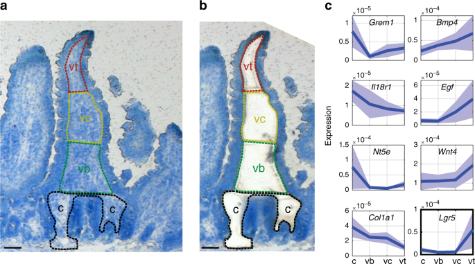
Lgr5 Telocytes Are A Signaling Source At The Intestinal Villus Tip Nature Communications
c. Renewal of the layers in contact with the villi which helps in absorption. 2. Propulsion: The chyme is moved over the large area of small intestine to facilitate digestion and absorption and the residues are propelled downwards to the ileocecal junction to reach the large intestine, mostly for excretion.

Draw A Schematic Diagram Of Villus In Small Intestine Explain How Digestive System

Villi Function Definition Structure Video Lesson Transcript Study Com
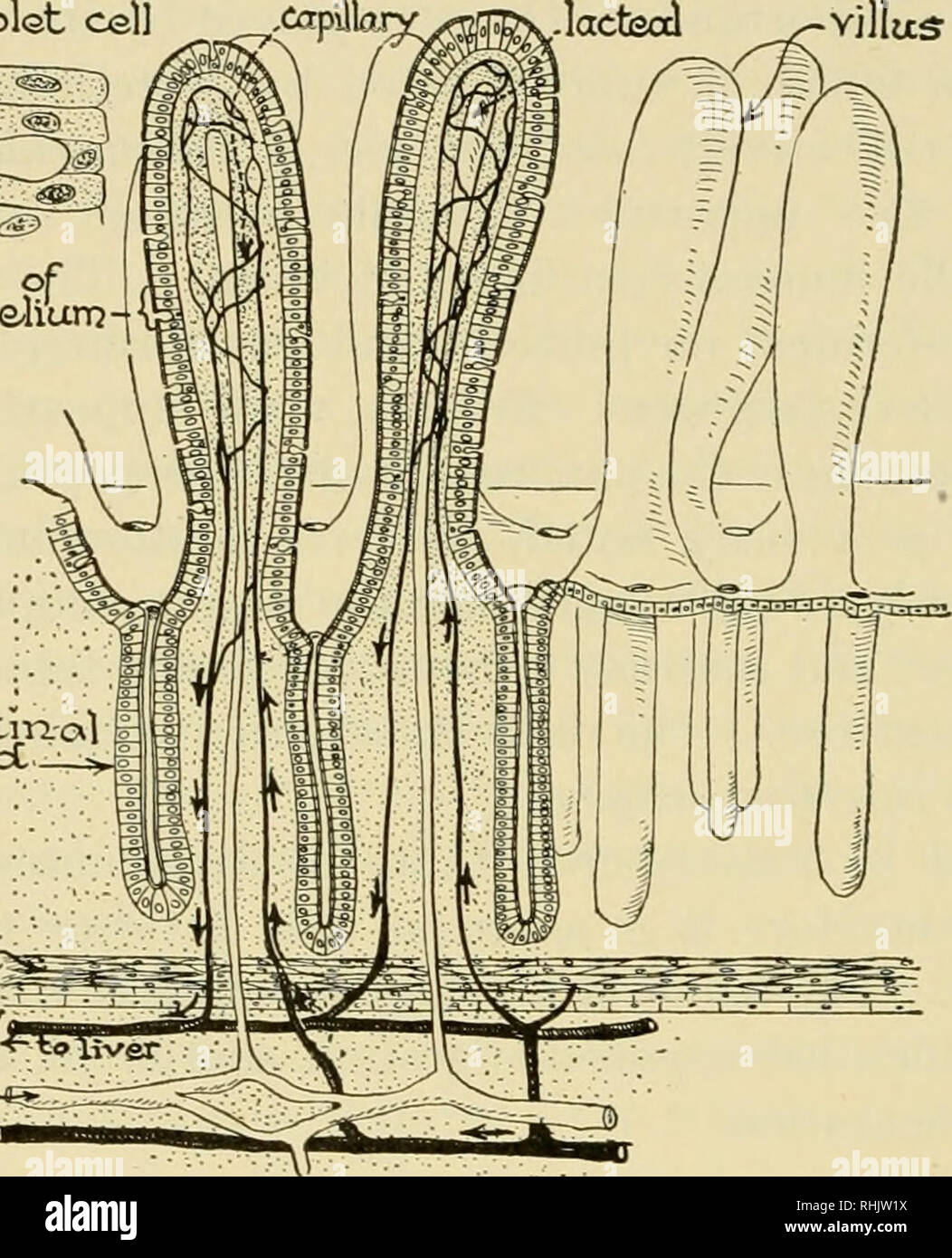
Biology The Story Of Living Things Cells Of Epithaliunj Intestm Al Glcxrjcc Niuscler Vein Icccteal Arter I Diagram Of Intestinal Villi And Glands Can You Explain The Part Played By The Villi In

The Diagram Given Below Represents A Section Of Small Intest
Digestive The Histology Guide

The Diagram Shows Part Of A Villus A Villus Is A Tiny Fingerlike Projection In The Inner Wall Of The Brainly In
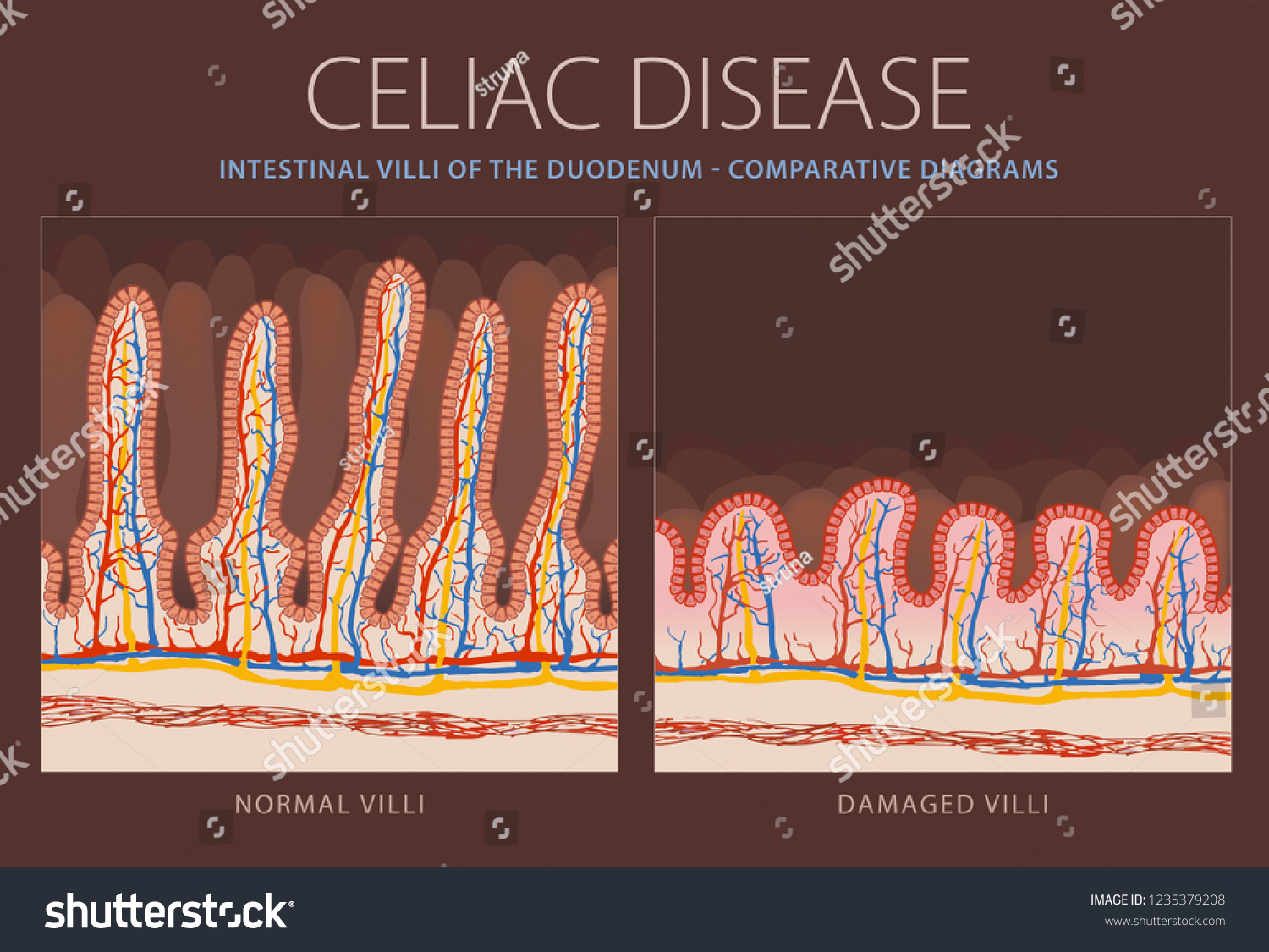
Healthy Intestinal Villi Damaged Villi Celiac Stock Vector Royalty Free 1235379208
Villi Illustration Twinkl

Villi Diagram Quizlet

Pin By Jessica Joyce On Systems Gastrointestinal Lymphatic System Anatomy And Physiology Lymphatic

An Animated Diagram Of Villi In The Smal Stock Video Pond5
1

The Given Diagram Shows The Internal Structure Of A Villus Label The Parts 1 To 4 Give The Function Of Brainly In

Portion Of A Villus Diagram Google Search Easy Doodle Art Teaching Practices Simple Doodles

183 Small Intestine Villi Stock Photos Pictures Royalty Free Images Istock
133 Intestinal Villi Vector Images Intestinal Villi Illustrations Depositphotos

Clear Diagram Enlarged View Of A Villus Brainly In
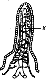
The Diagram Shows A Section Through A Villus What Is The Function Of Structure X Cbse Class 11 Biology Learn Cbse Forum


Comments
Post a Comment